
Lung imaging
Imaging lung function with hyperpolarized xenon-129
Xenon-129 magnetic resonance imaging shows lung function region by region, with no ionizing radiation.
What it shows
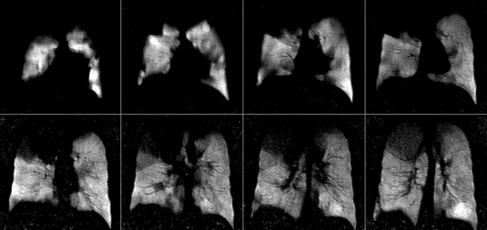
A conventional MRI scan shows almost nothing in the lungs, because the lungs contain mostly air. For a xenon scan, the patient inhales one or more breaths of hyperpolarized xenon-129. Some protocols use a single breath-hold, and others use free breathing. The scanner detects the xenon gas directly. The image shows regional ventilation: which parts of the lungs the gas reached, and which parts it did not. The resolution is about three millimeters.
Beyond ventilation: the dissolved phase
Xenon also dissolves in tissue and blood. It passes from the air spaces into the lung tissue, the blood plasma, and the red blood cells. Oxygen follows the same path. The xenon resonance has a different chemical shift in each of these compartments. One scan can therefore show how much gas reached the tissue and how much reached the red blood cells, in each region of the lungs. This is a direct image of gas exchange, the main function of the lungs. No other imaging method provides it.
Many researchers in the field now expect dissolved-phase imaging to become the most important clinical use. It is also the most demanding. Only one or two percent of the inhaled xenon is dissolved at any moment. The image must be formed from that small fraction of the signal. It requires highly polarized gas in sufficient quantity. Our polarizers were designed to deliver both.
Dissolved xenon also travels with the blood to other organs. Research groups have used hyperpolarized xenon to image the brain.
References: Mugler et al., Proceedings of the National Academy of Sciences 107, 21707 (2010); Qing et al., Journal of Magnetic Resonance Imaging 39, 346 (2014); and Ruppert et al., Scientific Reports 8, 7310 (2018), listed under Publications.
Clinical studies so far
MagniXene® is our hyperpolarized xenon-129. It has been studied in people under an Investigational New Drug application with the U.S. Food and Drug Administration since February 2005.
Clinical studies have been performed in healthy volunteers and in patients with asthma and chronic obstructive pulmonary disease. One published study compared xenon MRI with the current clinical standard, which is scintigraphy with an inhaled technetium-99m tracer. It enrolled fifty-nine subjects. It concluded that xenon MRI provides equivalent information. Xenon MRI also has higher spatial resolution, and it uses no ionizing radiation.
Reference: Peiffer et al., Academic Radiology 31, 1666 (2024), listed under Publications.
A double-blinded trial, at a different medical center, enrolled thirty patients with severe asthma. Xenon images were used to select the airways to treat by bronchial thermoplasty. The guided treatment took one session. The standard treatment takes three.
Reference: Hall et al., American Journal of Respiratory and Critical Care Medicine 202, 524 (2020), listed under Publications.
Guiding radiotherapy
Conformal radiotherapy irradiates a tumor from a wide range of directions and angles. The beams overlap at the tumor, where the dose accumulates to a lethal level. Each beam is shaped to spare as much healthy tissue as possible. Some harm to nearby healthy tissue is still unavoidable. It can reduce the patient’s exercise tolerance and quality of life after treatment. Some patients have lungs with marked functional heterogeneity, from advanced COPD or from earlier radiotherapy. For these patients, identifying and preserving the remaining healthy lung tissue could improve outcomes.
A regional map of xenon gas transfer to red blood cells shows which regions of the patient’s lungs function best. The treatment plan can then direct radiation dose away from those regions. This approach is called functional avoidance. It preserves lung function that the patient will need after treatment.
We believe this use is medically relevant and financially sustainable. The map changes a treatment decision. The patient is already being scanned for treatment planning. With partners at an academic medical center, we hold funding from the National Institutes of Health for a clinical trial of xenon-guided radiotherapy planning. The trial tests the hypothesis that functional-avoidance conformal radiotherapy improves outcomes for lung-cancer patients with functional heterogeneity.
Regulatory status
In the United States MagniXene® is an investigational drug. It is supplied for research under an Investigational New Drug application and is not approved by the FDA for clinical use. We give regulatory support to the investigators who use our polarizers.